

Collin & Kirk Optometrists has a proud belief that your vision deserves the best of service and care that we can offer.
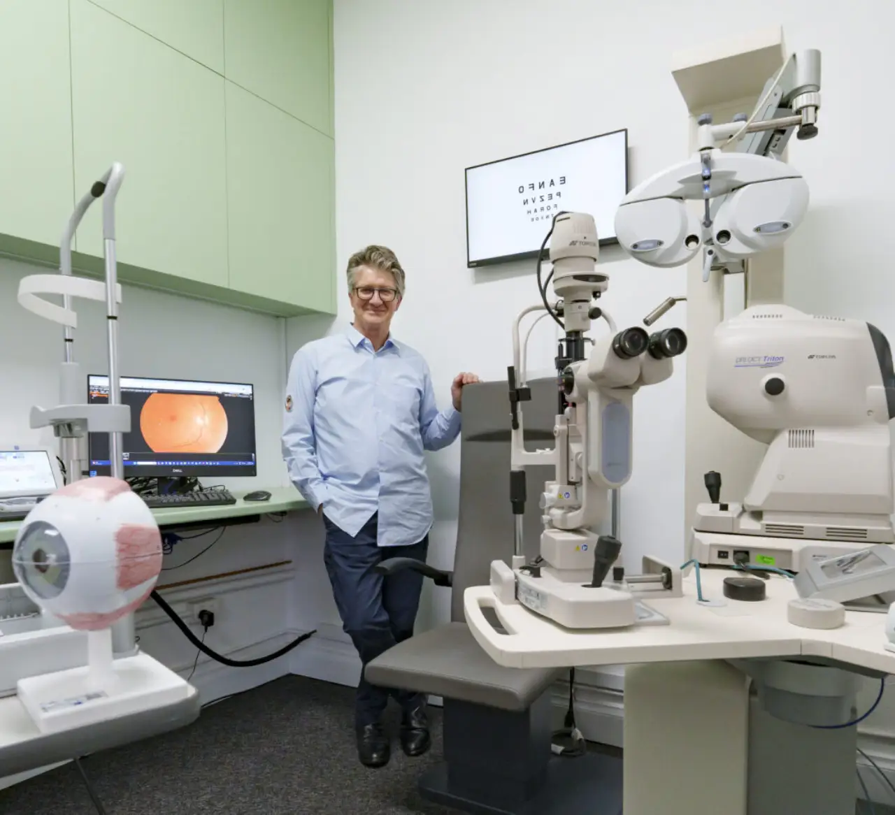
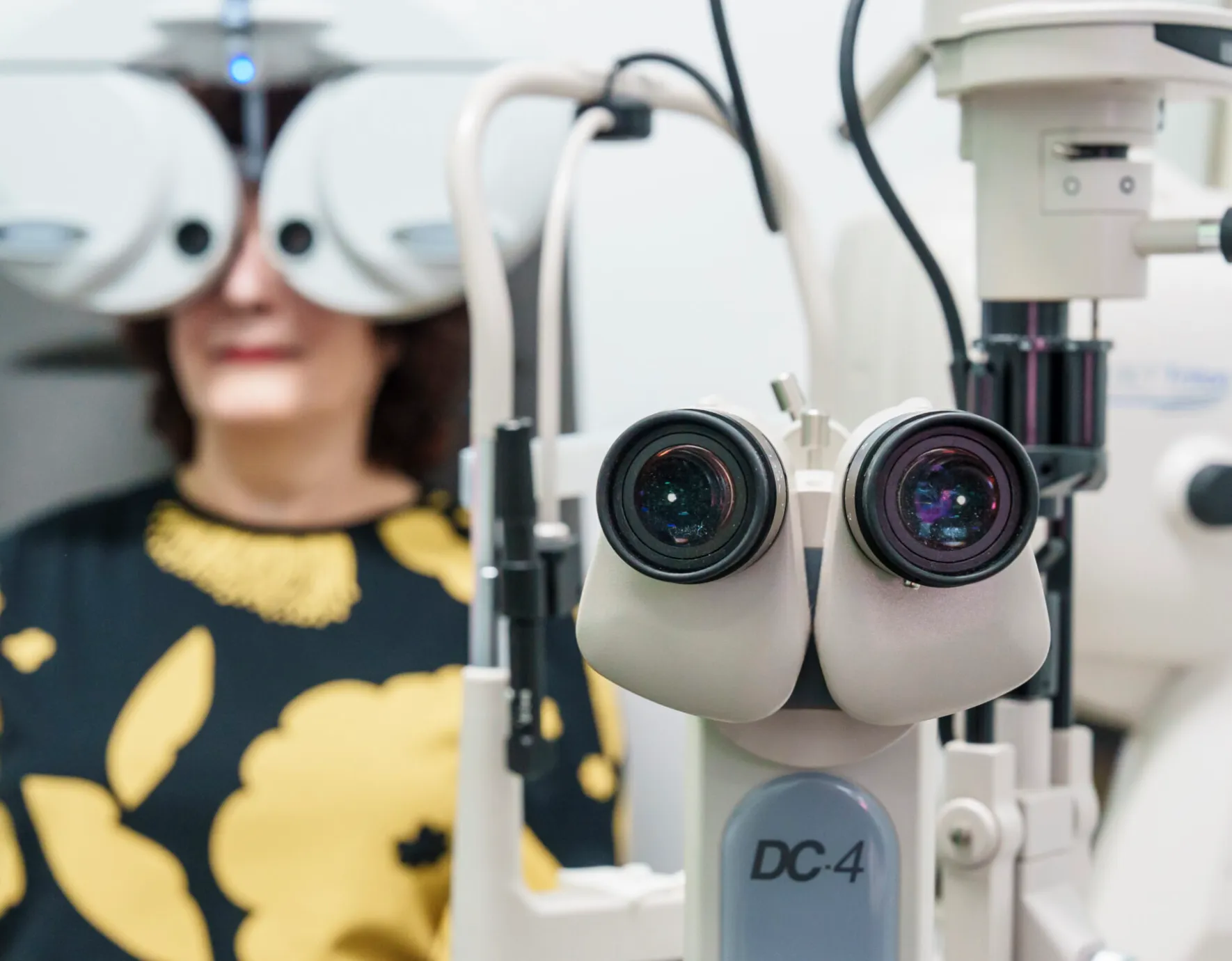
At Collin & Kirk Optometrists, we provide comprehensive eye examinations designed to assess more than just your prescription—we prioritise your long-term eye health. Using state-of-the-art diagnostic technology, we can detect early signs of eye conditions such as glaucoma, macular degeneration, and diabetic eye disease.
We recommend a comprehensive eye exam every two years.
Your initial eye examination
Your eyes are unique, and so is your vision care.
Our thorough eye exams go beyond checking your prescription – we assess your overall eye health, detect early signs of conditions like glaucoma and macular degeneration, and provide tailored recommendations to proetct your long term vision. Using state-of-the-art technology like the Rodenstock DNEye® Scanner, we ensure precise results for crystal-celar sight and comfort.
- Your eye examination may include:
- Digital imaging of the back of your eye
- A scan of your nerves and retina
- An assessment of the front of your eye
- A glaucoma check
- A check for macular degeneration
- Measurements of the shape and length of your eye
- A review of the movement and function of your eye
- A detailed assessment for spectacles or contact lenses
What you need to know
How often should I have an eye exam?
We recommend having an eye exam every two years, or more frequently if you have an existing eye condition, a strong prescription, or a family history of eye disease. Regular exams help detect vision changes and eye health issues early.
What happens during a comprehensive eye exam?
Our eye exams assess more than just your prescription. We check for signs of glaucoma, macular degeneration, and diabetic eye disease using advanced technology like the Rodenstock DNEye® Scanner. We also evaluate how your eyes work together, ensuring optimal vision and long-term eye health. Our personalised approach helps you achieve the clearest, most comfortable vision possible.
Do I need an eye exam if my vision seems fine?
Yes, even if your vision feels normal, an eye exam can detect hidden conditions such as glaucoma, early cataracts, or retinal diseases before symptoms appear. Some eye diseases develop gradually and can go unnoticed until significant damage occurs. A routine examination at our Thornbury clinic ensures early detection and prevention for lifelong eye health.
Can an eye exam detect other health conditions?
Yes, an eye exam can reveal signs of systemic conditions like diabetes, high blood pressure, and even neurological issues. Changes in blood vessels, optic nerves, or retinal health may indicate underlying health concerns. Our comprehensive assessments provide valuable insights into your overall well-being, helping you manage potential health risks early.
What should I bring to my eye exam?
Bring your current glasses or contact lenses, a list of any medications, and details of any eye or health conditions. If you have a referral or past eye test records, these can also be helpful.
Do you offer eye exams for children?
Yes, we provide eye exams for children of all ages. Early detection of vision problems is crucial for a child’s development and learning. We conduct a thorough exam to identify any issues with their vision, ensuring they get the right corrective treatment and avoid future complications.